Arteries supply the tissues in the Human body with oxygenrich blood Veins drain the same tissues Learn all about their microscopic structure on this full Histology Of Arteries Veins And Capillaries Preview Blood Vessels Lab Blue Histology Vascular System Artery Vein And Nerve Slide Peripheral Arteries Veins And Artery Blood Vessel Under Microscope View For Education Histology Top 60 Arteriole Stock Photos Pictures And Images IstockAbout Press Copyright Contact us Creators Advertise Developers Terms Privacy Policy & Safety How works Test new features Press Copyright Contact us Creators
Histology Slides 1
Artery vein nerve histology
Artery vein nerve histology- Histology of blood vessels 1 Blood Vessels 2 Objectives • Introduction • Components of circulatory system • Blood vessels – Basic structure • Arteries Elastic & Muscular • Arterioles, Metaarteriole • Capillaries Continuous, Fenestrated, Sinusoidal • Venules, Veins • Clinical CorrelationWe've gathered our favorite ideas for Artery And Vein Histology Slide, Explore our list of popular images of Artery And Vein Histology Slide and Download Photos Collection with high resolution


Human Artery Vein Nerve Cross Section Prepared Microscope Slide Eisco Labs
Arteries possess the all three basic histological layers of all cardiovascular tissues (See Cardiovascular Histology)The diameter and histological features of arteries transition throughout the cardiovascular system in accordance with their different functionalities, allowing for their categorization into three distinct histofunctional subtypes described further below Artery vein nerve under microscope リンクを取得 ;Arteries and veins of the lumbar nerve roots and cauda equina Dommisse GF, Grobler L An abundant vascular system supplies the nerve roots, within the cauda equina, in the spinal canal and in the nerve root tunnels The arterial supply of nervous tissue, as noted by previous observers, is "just adequate for its minimal needs" The veins of the area are classified into 3 groups, those of
» Histology Slides Print;Pulmonary arteries and veins These arteries have thin walls as a result of a significant reduction in both muscular and elastic elements, while the veins have a welldeveloped media of smooth muscle cells Umbilical vessels These arteries have two layers of smooth muscle cells without a prominent internal elastica or adventitia The vein hasFind the perfect artery and vein histology stock photo Huge collection, amazing choice, 100 million high quality, affordable RF and RM images No need to register, buy now!
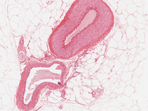
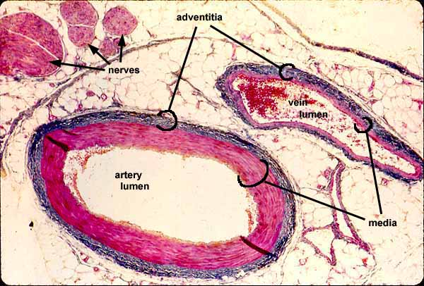
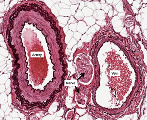
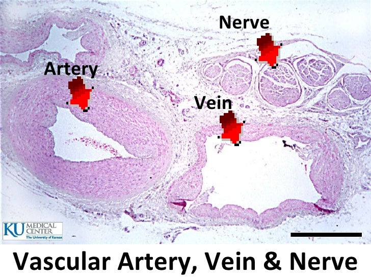
The main difference between nerve and vein is that nerve is an axon bundle of neurons in the peripheral nervous system, which carries nerve impulses whereas veins are blood vessels Artery, vein, nerve, trichrome stain Peripheral arteries, veins, and nerves tend to travel and branch in parallel Wherever one of these structures is found, the other two are likely to be closeby In this trichrome stained specimen, collagen is colored blue and smooth muscle is red Red blood cells in the venous lumen are brighter red The background is adipose connectiveSIU SOM Histology INTRO artery, vein, and nerve slide Peripheral arteries , veins , and nerves tend to travel and branch in Saved by SIU School of Medicine


Histology Slides 1


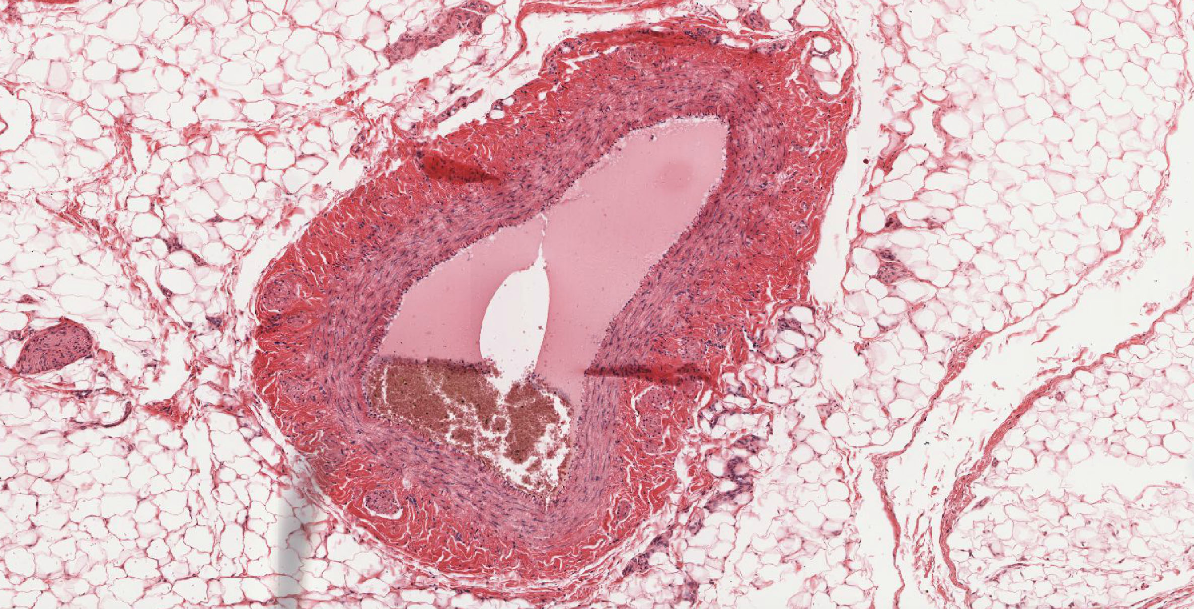
Muscular Artery Vein And Nerve Bundles Surrounded By Adipose Tissue The Artery Is Identified As Having
I will make it simple to identify any artery histology slide with proper identifying pointsTissue layer that contains veins, nerves, and Purkinje fibers b Myocardium consists oflayers ofcardiac muscle cells arranged in a spiral fashion about the heart's chambers and inserted into the fi brous skeleton The myocardium contracts to propel blood into arter ies for distribution to the body Specialized cardiac muscle cells in the atria produce several peptides including atrialMeyer's Histology Online Interactive Atlas and Virtual Microscope Improve your identification and understanding of histological structures!
:background_color(FFFFFF):format(jpeg)/images/library/3606/TIdQTQlRbloxtCUKcs2g_Arterioles.png)


Blood Vessels Histology And Clinical Aspects Kenhub
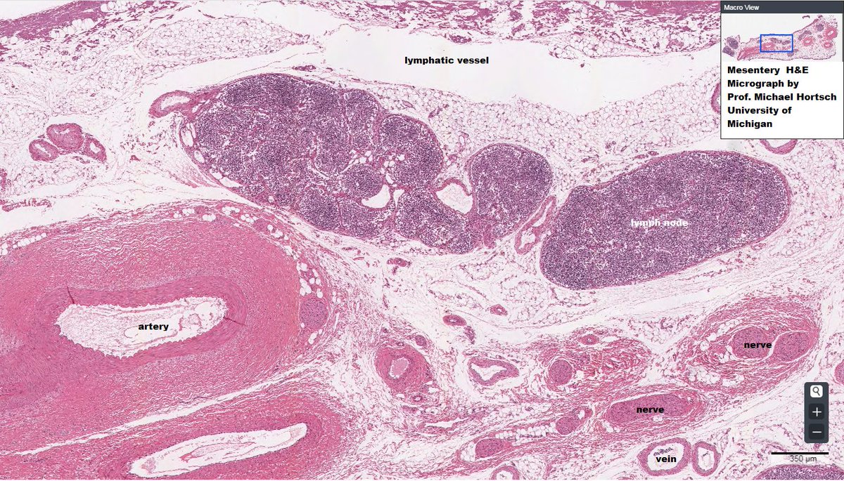


Twitter पर Dr Amanda J Meyer Is Double Pfizered Easy To Appreciate A Neurovascular Bundle In This Micrograph Artery Vein Lymph Nerve Anatomy Histology Vmdatabase T Co Lo8vq8dfjq
The blood vessels Venules The central vein is a small vein structurally characterised by its central position in the lobule of the liver functionally characterised by contribution to the larger whole but being the origin of the hepatic venous system that transports metabolically rich products to the rest of the body part of hepatic venous system made up from terminal branches of theView over over 4000 high resolution images of histological structures accompanied by interactive descriptive text that labels relevant histological details of every cell and tissue in the human body The Histology Guide Circulatory System Tissue Of Arteries And Veins Under The Microscopic Physiology Of A B Pancreases With Reliable Artery And Vein Anastomoses Had Gsc Go Science Crazy Mammal Artery Vein And Nerve Microscope Human Medium Sized Artery And Vein Microscope Slide Amazon Ca Mammal Artery Vein And Nerve C S 7 M Hematoxylin And Eosin Tissue Of Arteries And Veins
:background_color(FFFFFF):format(jpeg)/images/library/3607/bxosDBcdwLdq47dXkSy4A_Fenestrated_capillaries.png)


Blood Vessels Histology And Clinical Aspects Kenhub


Histology Of Umbilical Cord In Mammals Intechopen
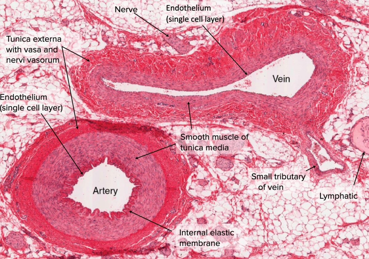
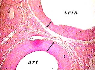
In larger arteries, elastic fibers unite to form a lamella which alternate with the layers of muscular fibers These fibers are connected with the membrane of the tunica intima In the largest arteries, there are a few bundles of white connective tissue that are wrapped around the tunica media Tunica adventitia The tunica adventitia is the outer layer of an artery, it surrounds the tunicaPeripheral nerve Venules continue into small veins, which possess a thin tunica media, composed of scattered smooth muscle, and a prominent tunica adventitia Like venules, veins also possess valves The wall of the small vein is much thinner and less regular than that of the small artery it accompanies 100xBasic Histology Artery and Vein You will often be able to distinguish arteries and veins, especally when they run together, as they usually do Arteries have thicker walls and tend to have narrower lumens They have to constrict and dilate to control how much blood flows where, and they must bear the powerful force generated by the heart Because of the large amount of


Histology Of Peripheral Nerves Anesthesia Key


Massasoit Instructure Com Courses Files Download Wrap 1
Histology This section of dentaljuce has over 400 histological slides, showing tissues from all organ systems in their healthy state Each tissue/organ slide set has an explanatory accompanying text which desribes its structure, function and role Histology Lab Image Information 1 Made by Luke Ausley (Feel free to edit) 2 Simple Squamous Epithelium Glomerulous Bowman's Capsule Vascular Artery, Vein & Nerve Nerve Artery Vein 33 Vascular Artery, Vein & Nerve Epithelial Tissue Part of the Circulatory system 34However, when examining the histology, the tunics in veins are not as clearly marked off as are the tunics in arteries Although all the three layers are present, there is less smooth muscle and connective tissue This makes the walls of veins thinner than those of arteries This is because blood in the veins has less pressure than in the arteries Because the walls of the veins are
:background_color(FFFFFF):format(jpeg)/images/article/en/histology-of-the-vascular-network/qTcfa6edhRn9fZVNzTw_aCakVCMk7M8xtaJDDC6A4w_Artery.png)


Blood Vessels Histology And Clinical Aspects Kenhub


General Histology Pptx د حارث Muhadharaty
Major arteries By definition, an artery is a vessel that conducts blood from the heart to the periphery All arteries carry oxygenated blood–except for the pulmonary arteryThe largest artery in the body is the aorta and it is divided into four parts ascending aorta, aortic arch, thoracic aorta, and abdominal aorta After receiving blood directly from the left ventricle of the heart, theArteries'and'Veins' ComparaveStructureof vein,'the'kidneys,'gonads'and'dorsal'por0ons'of'the' musculature''' '' '9In'fish,'the'system'is'modified'to'form'arenal'portal'system'' Blood'from'the'tail'and'posterior'trunk'flows'into'veins'called' renal'portal'veins'through'capillaries'along'the'HISTOLOGY LAB VII CARDIOVASCULAR SYSTEM BE ABLE TO IDENTIFY arteriole artery capillary cardiac muscle endo, myo, and epicardium elastic artery internal and external elastic laminae Purkinje fiber tunica intima, media, and adventitia vas vasi (pl vasa vasorum) vein venule Slide #170 (Artery, vein, and nerve) There are several features that allow one to distinguish between arteries
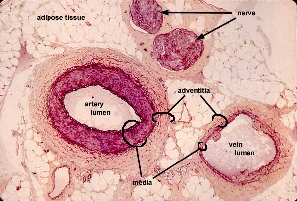


Biology 12 Gladstone Test Wednesday April 6 Let S Examine Specimens Update


Jerad Gardner Md Blood Vessels Nerves Usually Travel Alongside One Another This Section Of Normal Subcutis Shows A Nerve Vessel Weaving In And Out Of The Tissue Plane
View L03_Vessels_Arteries_Vein_Lymphatics_Nervespdf from BIOL 123 at King Saud bin Abdulaziz University for Health Sciences (Jeddah) Histology of Vessels Arteries, Vein, Optic nerve Identification points Innumerable transverse sections of myelinated (oligodendrocytes) axons of ganglion cells arranged in fascicles are seen In the middle central retinal artery and central retinal vein is seen At the periphery – the nerve is surrounded by three layers of meninges – duramater, arachnoidmater and piamaterIn the vein, the same 3 tunics can be seen but the tunica media is reduced, and the tunica adventitia is wider compared to the artery nervi vasorum (nervi vascularorum) vascular nerves that innervate both arteries and veins and control vessel vasodilation and vasoconstriction


Histology Diagrams General Histology Specific Points


Artery Vein And Nerve Histology A P1 Flashcards Quizlet
Start studying Artery, Vein & Nerve Histology Learn vocabulary, terms, and more with flashcards, games, and other study toolsArtery, Vein & Nerve 400x Arteries and veins are located throughout the body connecting to the heart They are part of the circulatory system that carries blood to and from the heart and other tissues and organs#113 Artery, vein and nerve, primate (H&E) Identify the cross section of peripheral nerve (The nervous tissue has shrunk within the perineurium during tissue processing) Only some of the axons within the nerve have been cut in true cross section What are the cells within the nerve whose nuclei are stained?
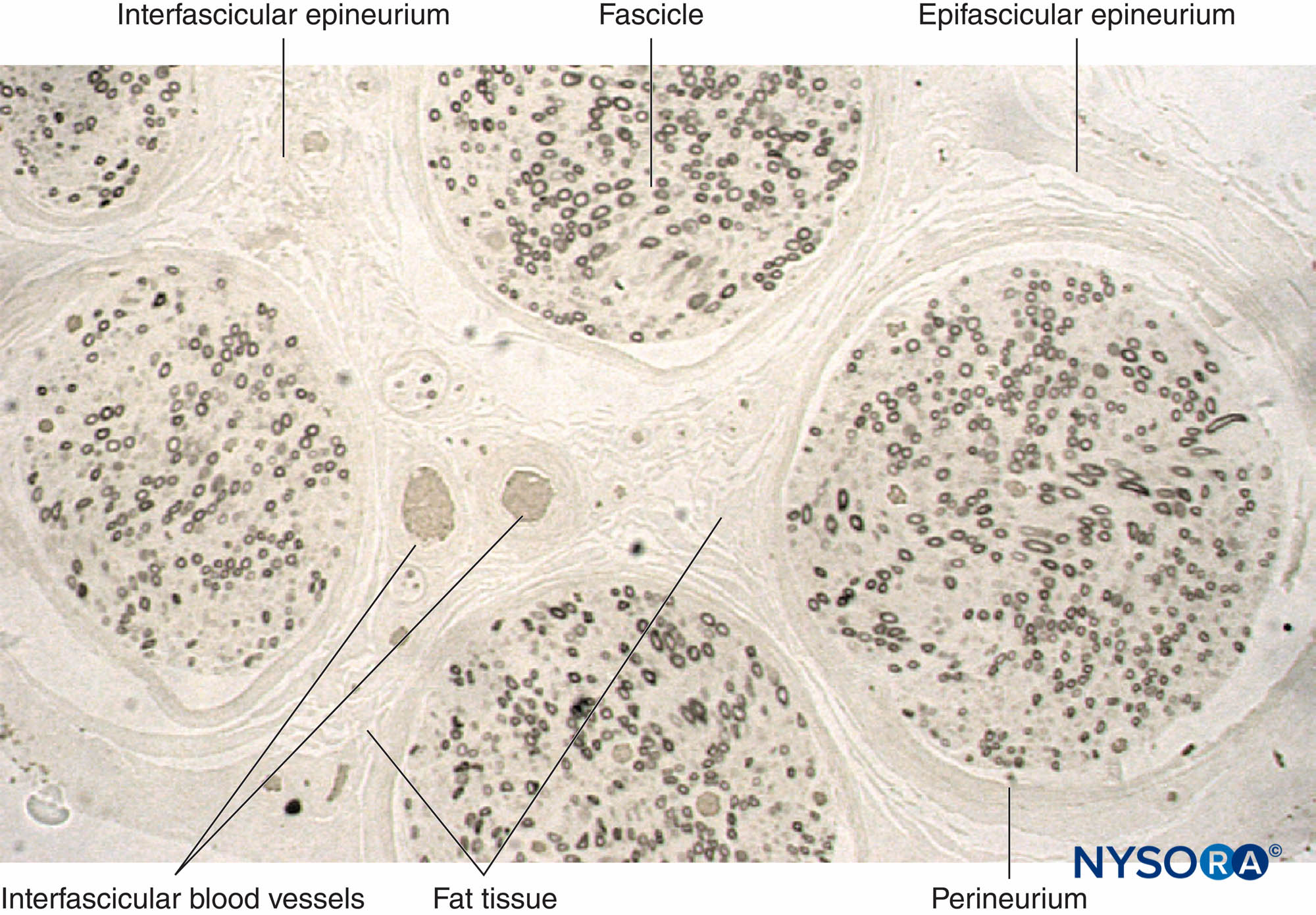


Histology Of The Peripheral Nerves And Light Microscopy Nysora


Human Artery Vein Nerve Cross Section Prepared Microscope Slide Eisco Labs
Arteries emanated from the base to the edge of the labia minora, where there was a larger feeding artery, and the arteries were anastomosed The veins formed anastomotic branches in the same direction as the edge of the labia minora Arteries and veins that accessed the labia minora were successfully perfused at the same time with no obvious association Sensory nerveImage Result For Artery And Vein Histology Arteries And Click Images to Large View Image Result For Artery And Vein Histology Arteries And Anat2511 Circulatory System Embryology Click Images to Large View Anat2511 Circulatory System Embryology Histology Slide Download Click Images to Large View Histology Slide Download Mammal Artery Vein And Nerve Cs 7 µmThe arteries adjacent to the lesser saphenous vein and sural nerve were investigated in 10 fresh cadavers that had been systemically injected with a lead oxidegelatin mixture The accompanying arteries were found to lie along the lesser saphenous vein and sural nerve and to nourish the skin through venocutaneous and neurocutaneous perforators On the basis of the anatomy of these


Artery And Vein Digital Slide Youtube


Untitled Document
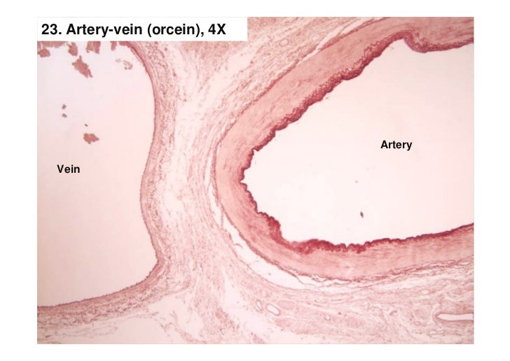
Artery, vein, nerve, elastin stain Peripheral arteries, veins, and nerves tend to travel and branch in parallel Wherever one of these structures is found, the other two are likely to be closeby In this specimen, elastic tissue is colored dark brown, collagen is pale pink and cytoplasm (in smooth muscle and nerve) is purple The background is adipose connective tissueMediumSized Artery And Vein Plate 103 Arterioles, Capillaries, And Lymphatic Vessels Plate 104 The Heart The Wall Of The Atrium Overview The human body is a wellconstructed collection of trillions of cells As indicated in Chapter 1, each of those cells needs essential raw materials such as food, water, and oxygen to survive Because most of the body's cells are distant from foodArtery and vein histology labeled Google Search


Histology Slides For The Cardiovascular System Slides Are Presented In Order Of Magnification As You View The Following Slides Make Sure You Can Accomplish Ppt Download


Animal Organs Cardiovascular System Atlas Of Plant And Animal Histology
#113 Artery, vein and nerve H&E Open with WebViewer All blood vessels are lined by simple squamous epithelium called the endothelium Examine the endothelial lining of the large artery and vein At the level of the light microscope the cytoplasm is difficult to visualize the lining cells, however nuclei can be distinguished In the large muscular artery there is a layer of highlyStart studying blood vessel histology Learn vocabulary, terms, and more with flashcards, games, and other study tools Start studying Artery, Vein & Nerve Histology Learn vocabulary, terms, and more with flashcards, games, and other study toolsThe veins are originated in the cortex and follow parallel to the interlobular, arcuate, interlobar, and segmental arteries until forming the main renal vein Figure 1 The arcuate arteries (green arrow) run by the interstitial space between the cortex


Artery Vein Nerve Histology Diagram Quizlet


Arteries
The lateral cutaneous nerve of the forearm descended deeply along the cephalic vein in 124 cases (97 %), while the medial cutaneous nerve of the forearm descended superficially along the basilic vein in 94 (73 %) A superficial brachial artery was found in 27 cases (21 %) and passed deeply under the ulnar side of the MCV A median superficial antebrachial artery was found in 1 case (1 Blood vessel histology Author Lorenzo Crumbie MBBS, BSc • Reviewer Dimitrios Mytilinaios MD, PhD Last reviewed Reading time 16 minutes It would be impossible to get blood to the predestined locations without the vascular pathways Blood vessels form the extensive networks by which blood leaves the heart to supply tissue Additionally, other blood vesselsFind a small nerve elsewhere in the
:background_color(FFFFFF):format(jpeg)/images/library/3615/NOpbXd2vHcY0t8xD0mZLQ_Vasa_vasorum.png)


Blood Vessels Histology And Clinical Aspects Kenhub


Cardiovascular System Histology
Mammal Aorta, Artery, Veins Microscope Slides 3 Items $630 $765 Qty Discount Available View Details From cat or dog Stained to show general structures Sort by Mammal Aorta cs 7 µm Verhoeff's stain Microscope Slide Item # $765 Qty In Stock Quick View Mammal Artery & Vein cs 7 µm H&E Microscope Slide Item #3140 $645 QtyHistology flashcards The male reproductive system Learn all organs, nerves, arteries, veins and tissues on the go (Kenhub Flashcards Book 7) eBook Kenhub, Kenhub Amazoncouk Kindle Store9月 09, 21 When the SCA originates from the basilar artery, it passes below the third cranial nerve, and when it comes from P1, it can pass above the oculomotor nerve Classically, the SCA transits under the third cranial nerve and the fourth cranial nerve


Mammal Nerve Transverse Section 64x Nerves Mammals Mammals Nervous System Other Systems Comparative Anatomy Of Vertebrates Animal Histology Photos


Venous Organization In The Transverse Foramen Dissection Histology And Magnetic Resonance Imaging In Journal Of Neurosurgery Volume 123 Issue 1 15 Journals
Histology of blood vessels Dr Calum Worsley and Associate Professor Donna D'Souza et al Blood vessels, namely arteries and veins, are composed of endothelial cells, smooth muscle cells and extracellular matrix (including collagen and elastin) These are arranged into three concentric layers (or tunicae) intima, media and adventitia Artery histology Arteries are the thickwalled blood vessels that carry blood from the heart to the capillaries You may ask to identify the three different types of arteries slides (large, medium, and small) at the histology laboratory No worries;In this study serial sections of the central retinal vein and artery in the anterior optic nerve from six globes (five from cornea donors and one exenteration specimen) were examined by image analysis and threedimensional reconstruction to determine their luminal characteristics The retinal artery was found to have a uniform perimetric length and crosssectional area The vein, however, had


Peripheral Nerve Histology Draw It To Know It


Description


Histology Slide Download Magscope Com
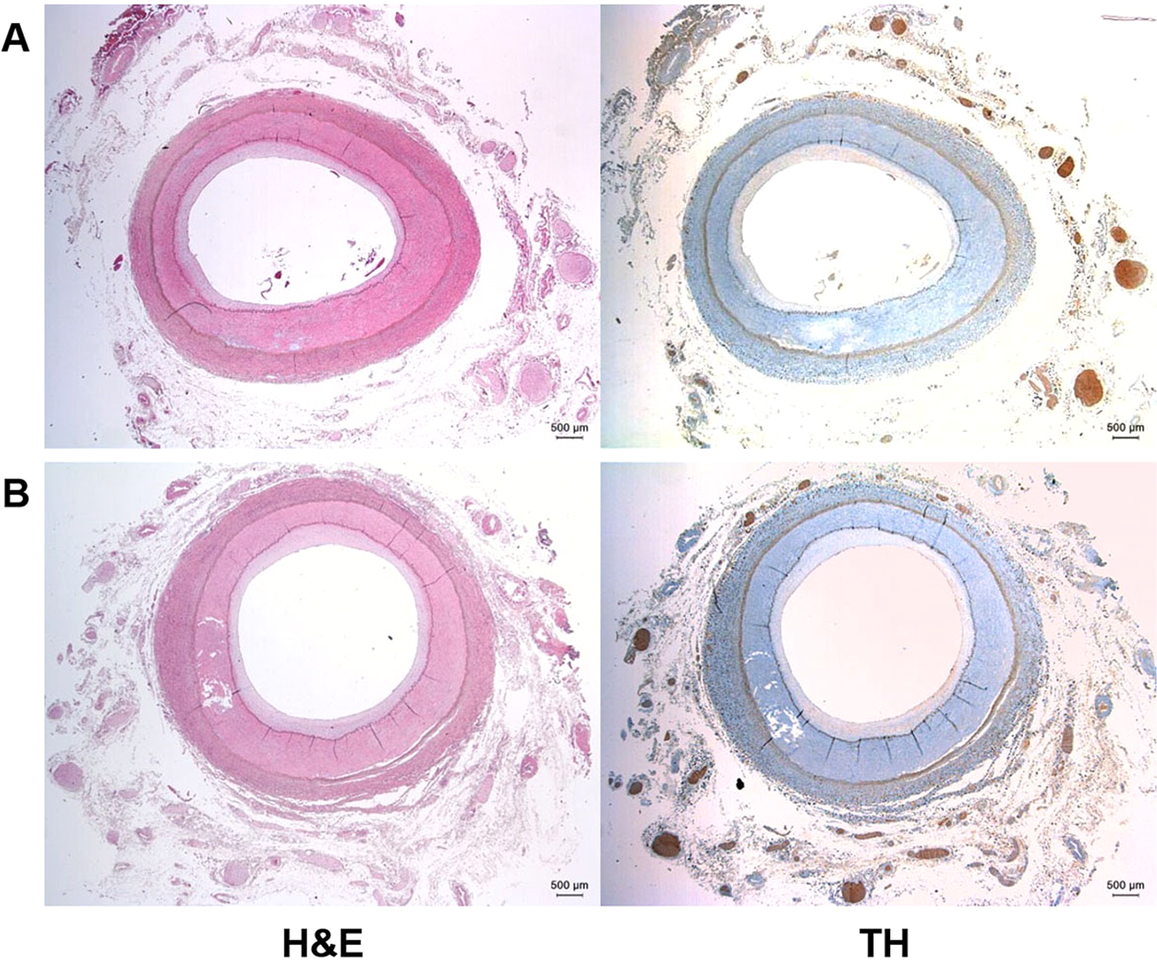


Anatomic Conformation Of Renal Sympathetic Nerve Fibers In Living Human Tissues Scientific Reports
:background_color(FFFFFF):format(jpeg)/images/library/3613/OgR38mkJaHfLSb8S17ENg_Lymphatic_vessel_of_the_dermis.png)


Blood Vessels Histology And Clinical Aspects Kenhub


Histology Laboratory Manual


A Novel Rat Microsurgical Model To Study The Immunological Characteristics Of Male Genital Tissue In The Context Of Penile Transplantation Fidder Transplant International Wiley Online Library


Cardiovascular System Histology


Histology Of The Retromolar Canal A Z Artery With A Thick Wall N Z Download Scientific Diagram


Capillary


Histology At Siu
:background_color(FFFFFF):format(jpeg)/images/library/13219/arteries-and-veins.psb_english.jpg)


Blood Vessels Histology And Clinical Aspects Kenhub


Histology Review Nervous Tissue Dr Tim Ballard Department


Histology Arteries And Veins Www Topsimages Com


Cardiovascular


Histology Slides For The Cardiovascular System Slides Are


Pin On Histology Nervous System


Artery Vein Nerve Histology Diagram Quizlet


Description


Block3 Fig 11 93w4046 Artery Vein Nerve Elastic Tissue Cs V E


Arteries


Human Structure Virtual Microscopy


Blood Vessel Color Images


Circulatory Np Histology


Peripheral Nerve Histology Histology Flashcards Draw It To Know It


Untitled Document
:background_color(FFFFFF):format(jpeg)/images/library/3608/jvqXu9FdCLcYYymIaZRwA_Sinusoidal_capillary.png)


Blood Vessels Histology And Clinical Aspects Kenhub


11 Circulatory System Ideas Circulatory System Arteries And Veins Anatomy And Physiology


Circulatory Np Histology


Erect Anatomy Illustration Of Male Erection Physiology With Cavernous Veins Arteries And Nerves Canstock


A Histology Of Common Hepatic Artery Day 90 Movat S Pentachrome Download Scientific Diagram


Nerve Tissue Al Maarefa College Nerve Tissue Cells Have Very High Ability To Respond To Stimuli Transmit Impulses Ppt Download


Lab 2 Epithelial Tissue Histology


Histology Of The Circulatory System The Cardiovascular System


Blood Vessels Artery Vein Nerve


Arteries Concise Medical Knowledge
:background_color(FFFFFF):format(jpeg)/images/library/3605/qqhmVxIKd7pxeygeKSM3xw_Muscular_artery_-_Coronary_artery__1_.png)


Blood Vessels Histology And Clinical Aspects Kenhub


Histology Page


Histology Of Umbilical Cord In Mammals Intechopen


Histology Of Blood Vessels


Artery Vein Nerve Prepared Microscope Slide 75x25mm Eisco Labs


Blood Vessels Lab


Http Www Phlebology Org Wp Content Uploads 15 03 01 Anatomy Embryology Histology Macroscopic Anatomy Embryology Kalva Pdf
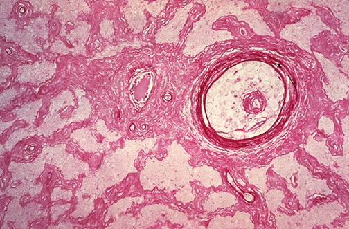


Ophthalmic Pathology


Muscular Artery Vein And Nerve Bundles Surrounded By Adipose Tissue The Artery Is Identified As Having A Thicker Wall And A Smaller Diameter Than Th Stock Photo Alamy


Artery And Vein C S


Mammal Artery Vein And Nerve Simple Squamous Annotated 40x Histology


Moran Core Retina Rpe Histopathology


Blood Vessels Lab


Cardiovascular System Histology


Nervous Tissue Ii


Blood Vessels Lab
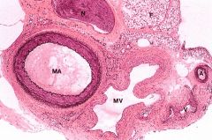


Cp Histology Anatomy Gu Som Flashcards Cram Com


Histological Section Of The Human Retina Retinal Vessels Download Scientific Diagram


Arteries


Arteries
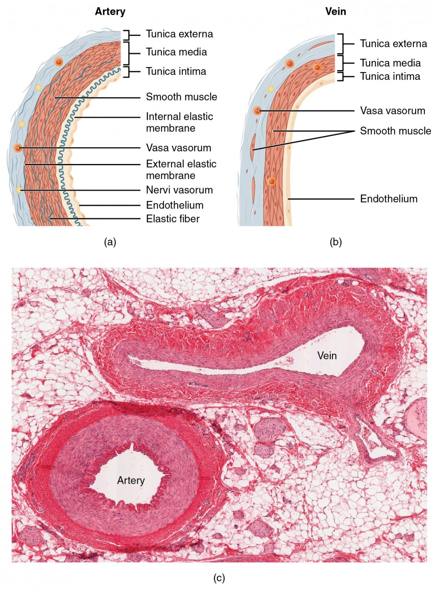


Structure And Function Of Blood Vessels Anatomy And Physiology Ii


Histology Of Blood Vessels


Untitled Document


Artery Vein And Nerve Histology A P1 Flashcards Quizlet


Histology Vessels


Large
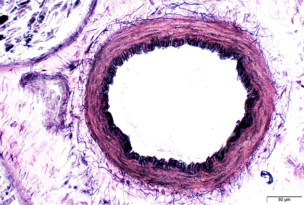


Vasculitis


Vetmed Tamu Edu Peer Wp Content Uploads Sites 72 04 9 Blood And Lymph Vessels Pdf
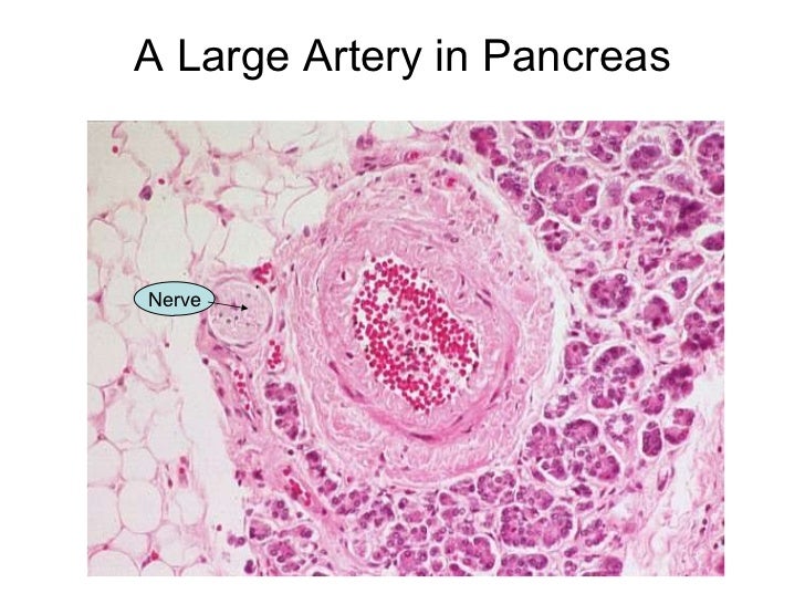


Basic Histology


11 Circulatory System Ideas Circulatory System Arteries And Veins Anatomy And Physiology


Optic Nerve Histology Youtube


1


Artery And Vein


1


Histology Of A Portal Triad Bile Duct Ha Hepatic Artery Pv Download Scientific Diagram


Blood Vessels Knowledge Amboss


Artery Vein And Nerve Histological Slide Youtube


Histology Lab Image Information


Small Veins And Muscular Arteries


Pin On Medical Laboratory Science


Arteries


Cardiovascular


0 件のコメント:
コメントを投稿